Cryo-electron Microscopy (Cryo-EM)
Including the W. M. Keck Center for Virus Imaging in BSL-3-
Introduction
The SCSB Cryo-electron Microscopy (Cryo-EM) Laboratory, located on the first floor of the Medical Research Building, features a BSL-3 containment for viral and pathogen work. The open research space (separate from BSL-3 containment) is designed for studying the structures of macromolecules, their complexes, cell organelles, and other biological systems using various EM techniques, including cryo-electron microscopy and (cryo-) electron tomography. The laboratory has three modern microscopes that are equipped for (cryo-) electron microscopy and (cryo-) electron tomography with automated data collection incorporated.
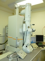
The 300 kV Thermo-Fisher Titan Krios cryo-EM is a state of the art ultra-high-resolution microscope with post-column electron energy filter and field emission gun (FEG), which is the brightest electron source currently available. This fully automated instrument yields near-atomic resolution images of biological macromolecules and their complexes and is capable collecting data without user intervention.
The high-resolution 200 keV JEM2200FS is located in the W. M. Keck Center for virus imaging in BSL-3 containment and permits the safe structural imaging of highly infectious pathogens that could not studied in the open research area. This is the first cryo-EM facility in the US designed for high-resolution structural studies of wild type infectious agents. The microscope can be controlled remotely through a computer network, which provides access for remote online users and largely extends our user base.
The JEM2100 is available for imaging of non-pathogenic specimens and negatively stained samples. All microscopes in the Laboratory are equipped for (cryo-) electron tomography using automated data collection procedure. The JEM2100 is used for both user training and structural studies. A shared Crystallographic/EM Computational Lab provides high-throughput image-processing with a dedicated 120-core EM Image-Processing Cluster. Images are stored in a local EMEN database that is archived remotely.
-
Instruments


Titan Krios JEM2200FS JEM2200BSL3 in the
W. M. Keck Center
for Virus ImagingJEM2100 -
Documentation
-
Contact
For more information please contact the SCSBMB manager.
Manager
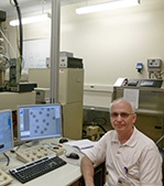
Michael Sherman, PhD
E-mail: mbsherma@utmb.edu
Tel: (409) 772-6310Research Scientist
Michael Woodson, PhD
E-mail: miwoodso@utmb.edu
Tel: (409) 772-6327Advisory Committee
- Scott C. Weaver, PhD
- Mark A. White, PhD, Manager, X-ray Crystallography Laboratory
- Y. Whitney Yin, PhD
-
Microscope Scheduling
New users and current users who have new research projects and/or new grants are asked to please complete the Usage form:
To schedule experiments, please submit Schedule Request Form:
- Schedule Request Form (NEW Webform)
- Instructions for Schedule Request Form
Instrument Scheduling
-
People
Misha Sherman, PhD 
Matthieu Gagnon, PhD 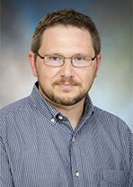
Petr Leiman, PhD 
Gabrielle Rudeno, PhD 
Thomas Smith, PhD 
Scott Weaver, PhD 
Y. Whitney Yin, PhD 
Michael Woodson, PhD
